This article is about explaination on the process of digestion in human beings with labelled diagrams, helpful for class 7, 8, 9 and class 10 biology (zoology) students.
Explaination on the process of digestion in human beings
The well described article on the process of digestion in human beings with lebelled diagram will be very much helpful for the NCERT and State board students of Class 6, 7, 8, 9, and class 10.
What is Digestion in Human Beings?
The process of digestion involves breaking down the complex chemical components in food into simpler forms that the body can easily absorb and use for development, energy, and repair.
It starts in the mouth and ends in the small intestine, involving a variety of organs (such as Mouth, Pharynx, Esophagus, Stomach, Duodenum, Colon, Liver, Gallbladder, Pancrease etc), enzymes, and chemical processes.
Understanding digestion entails investigating its stages, mechanics, and the role of each digestive component in the entire process.
Human digestion is a complex and intriguing trip that begins when food enters the mouth and finishes when nutrients are absorbed into the bloodstream.
This complex process comprises multiple organs and phases, each having a specialised job in ensuring the body receives the nutrients it requires for growth, energy, development, and repair.
- Explaination on the process of digestion in human beings
- What is Digestion in Human Beings?
- Step By Step Overview on the Process of Digestion in Human Beings
- Labelled Diagram of Human Digestive System For Class Notes
- Role Of Salivary Secretion In The Process Of Digestion of Human Beings
- Role Of Mouth and Oesophagus In The Process Of Digestion
- Role and Functions of Stomach in the Process Digestion
- Role and Functions of Pancreas in the Process Digestion
- Functions of Liver in Human Digestive Process
- Process of Digestion in the Small Intestine
- Role of Large Intestine or Colon

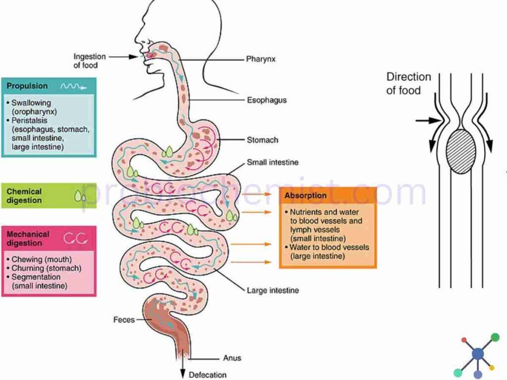
Step By Step Overview on the Process of Digestion in Human Beings
Digestive System performs different roles in our body, such as:
1. Process of Ingestion
Food is first chewed with the help of teeth, and then the chewed food is combined with saliva in the mouth to start the process of digestion.
Enzymes such as amylase which is found in salivary gland, found inside the buccal cavity, begins the breakdown of carbohydrates. The food materials which are chewed inside the mouth is called as “bolus”.
2. Swallowing of Food Materials
The chewed food, now referred to as the “bolus”, is ingested and passes down the oesophagus via a motionless, synchronised contraction of muscles known as peristalsis movement.
3. Digestion of Food Materials inside Stomach
Once the bolus enters the stomach, then the bolus is combined with the digestive juices. These juices have pepsin and hydrochloric acid, which are enzymes that begin to break down amino acids molecules and proteins.
Food is turned into chyme, a semi-liquid material, via the stomach’s churning process.
4. Digestion in Small Intestine (Duodenum, Jejunum and Ileum)
– Role of Digestion in Duodenum: The duodenum, the first segment of the small intestine, is where chyme next goes. It combines with pancreatic enzymes and liver bile at this point.
Pancreatic enzymes, such as Amylase, Protease and Lipase further helps in breaks down of proteins, carbohydrades, lipids, and fats. Bile juice helps in emulsification of fats.
– Role of Digestion in Jejunum: Nutrients enter the circulation in the jejunum and ileum (the remaining sections of the small intestine) through the intestinal lining.
Villi and microvilli, which resemble tiny fingers projecting from the inner walls of the small intestine. This small structures increases surface area of the small intestine and thus helps in absorption of food materials, nutrients.
5. Absorption and Transport of Nutrients
The wall present inside the gut or the intestine allows nutrients to enter the circulation or lymphatic system. These nutrients are carried by the blood to the liver, where they are processed and then distributed throughout the body for further process.
6. Process of Digestion in Large Intestine
The large intestine receives the leftover undigested food and waste materials.
Large Intestine is the place, where the Water and electrolytes gets absorbed, and waste material solidifies into faeces
7. Elimination of Metabolic Food Waste
The solid waste is held in the rectum until it is ready to exit the body via the anus during faeces.
This entire process guarantees that your body receives the nutrition it requires for proper functioning, growth and development of various organs while disposing of waste items.
Each step requires a combination of enzymes and other chemicals to break down and absorb the food you consume.
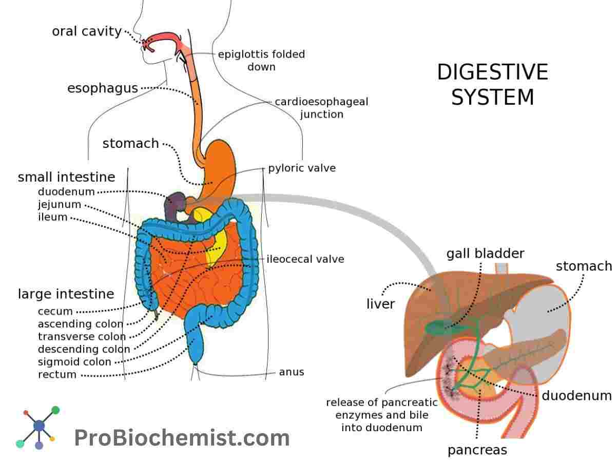
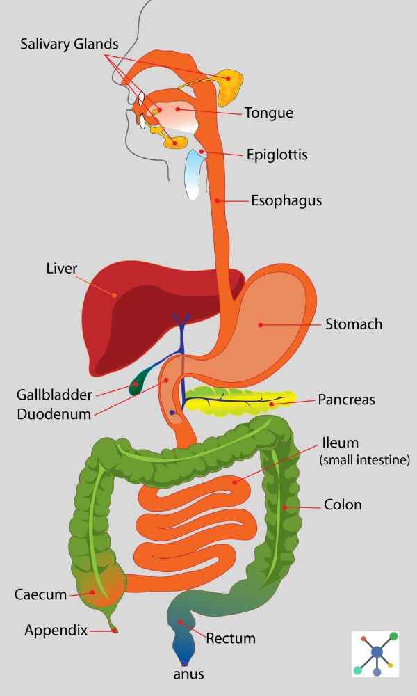
Labelled Diagram of Human Digestive System For Class Notes
Also Read: How To Make Human Digestive System Model – 3D Model Project for School, College and University
Role Of Salivary Secretion In The Process Of Digestion of Human Beings
There are 3 pairs of salivary glands –
- Parotid
- Submandibular (or submaxillary)
- Sublingual salivary gland.
Salivary glands contain chiefly two types of cells: ‘mucous’ cells and ‘serous’ cells. Both sets of glands contain their secretory cells in acini where the cells are arranged around a central lumen, which leads into a duct.
These ducts join to form intralobular and interlobar ducts which lead into the main duct.
All 3 pairs of salivary glands are supplied by ‘efferent’ and ‘afferent’ nerves.
Composition of Saliva
Daily Secretion: Average 1500 mL/ day.
Digestive Enzymes present in Salivary Secretion:
(i) ptyalin or salivary amylase
(ii) lysozymes (bactericidal)
(iii) kallikrein, a proteolytic enzyme
(iv) lipase, a lipolytic enzyme (secreted by glands on
the tongue, therefore, also called as lingual lipase).
Function of Salivary Gland in Digestive Process of Human Beings
- 1. Ptyalin (salivary α-amylase):
Ptylin helps in digestion of starch to 1 : 4α linkages producing α-limiting dextrin, and maltose (to some extent). It can only digest starch after the natural plant granules have been burst e.g. by cooking.
It acts in a neutral or faintly acidic medium (optimally at pH 6.5). Given time, it can digest starch to maltose.
Such digestion continues in the interior of the bolus of food formed by chewing and mixing with the saliva even when this bolus has reached the stomach.
Amylase digestion can thus continue in the stomach for approx. half an hour, until it is arrested by the excessive acidity of the gastric contents. Amylase is readily inactivated at pH 4.0 .
- 2. Mucin
(I) It lubricates the food, thus assists mastication and facilitates swallowing of food materials.
(II) It protects the oral mucosa.
(III) It aids speech by facilitating movements of lips and tongue.
- 3. It keeps the mouth moist and serves as a solvent for the molecules that stimulate the taste buds.
- 4. It minimizes risk of buccal infection and dental caries.
- 5. Buffers and ‘proline’ rich proteins in saliva helps to bind toxic tannins and maintain the oral pH at 7.0. At this pH saliva is saturated with calcium, therefore, teeth do not lose calcium to oral fluids. Thus it protects the tooth enameI. Acidic oral pH causes calcium loss from the teeth. Buffers in saliva also helps to neutralize gastric acid and relieve heart burn when gastrjc juice is regurgitated into the oesophagus.
- It is a vehicle for the excretion of certain drugs e.g. alcohol,and morphine; and of certain inorganic ions, such as potassium, iodine and thiocyanate etc.
Mechanism: Salivary Secretion by Salivary Glands
1. The acinar cells secrete K+ and HC03- by active process into the acinar lumen, accompanied by sufficient Cl– to preserve electrical neutrality.
Simultaneously, passage of water into the acinar lumen makes this primany secretion isotonic.
The salivary duct cells which drain the acini have a rich blood supply, therefore, they actively reabsorb Na+ and an accompanying anion (CI–) and transfer some K+ and HC03-into the saliva.
The duct cells being relatively impermeable to water make the final salivary secretion – hypotonic. Following secretion, the number of granules in the protoplasm of secreting cells falls sharply.
2. Saliva is a hypo-osmolar (hypotonic) secretion of salivary glands, therefore, metabolic activity i.e. Oxygen consumption of the gland is increased by 5 folds during secretory activity as compared to that at rest.
3. At rest, saliva contains more of K+ and less of Na+, Cl– and HCO3- as compared to their plasma concentrations.
As salivary flow increases, there is less time for ions exchange in the ducts , as a result saliva becomes less hypotonic (almost isotonic i.e. resembles the primary secretion. Na+, Cl– and HC03- concentration increases and K+ concentration decreases.
- Na+ concentration increases to a plateau concentration of 80-90 mEq/ L, and
- Cl- concentration increases to 50 mEq/ L. Na+ and Cl- concentrations of saliva are always lower than that in the plasma.
- HC03- concentration increases and exceeds that in the plasma, and
- K+ concentration decreases to 15-20 mEq/L. As a result of this, pH of saliva which is < 7.0 at low secretory rates increases to approx. 8.0 as the rate of salivary secretion increases.
4. Iodide and thiocyanate, which are secreted in saliva, are also actively transported from plasma directly into the lumen of the ducts by the cells of ducts wall. They are not transported by the acinar cells.
5. Aldosterone increases the Potassium (K+) concentration and decreases Sodium (Na+) concentration of saliva. (Aldosterone acts on the ductal cells, an action similar to its action on the kidneys).
Thus, a high salivary Na+ / K+ ratio is seen when aldosterone is deficient (Addison’sdisease).
Role Of Mouth and Oesophagus In The Process Of Digestion

Voluntary Oral Stage of Digestion : Role of Mouth in Digestion
- After mastication chewed food, moistened and lubricated by saliva, is rolled into a bolus; by movements of the cheeks and tongue the bolus comes to lie in the curve of the tongue. ·
- Contraction of the front part of the tongue presses the bolus against hard palate, while a series of movements of the middle part push it to the back, forcing it past the anterior pillar of the fauces (palato-glossal folds) to the root of the tongue.
- Swallowing commences by closure of the mouth and voluntary contraction of mylohyoid muscle,
which throws the bolus back between the anterior and posterior pillars of Jnuces (i.e. palato-glossal and palato-pharyngeal folds) on to the posterior pharyngeal wall. - Swallowing (Deglutition) Reflex in The Process Of Digestion In Human Beings:
It is triggered by afferent impulses arising from:
(i) mucous membrane covering anterior and posterior
pillars of fauces, and tonsils.
(ii) posterior pharyngeal wall
(iii) soft palate; and
(iv) epiglottis
These impulses travel in trigeminal (V) , glossopharyngeal (IX) and vagus (X) nerves to stimulate a group of-nerve cells located in the floor of IVth ventricle near the respiratory centre in the medulla. The efferent fibers pass through motor fibers of V, Vil (facial), IX, X and Xll (hypoglossal) nerves to the pharyngeal musculature and the tongue. This initiates involuntary pharyngeal stage.
Involuntary Pharygeal Stage: Process Of Digestion In Human
This stage shows involuntary, complex, closely coordinated movements in the pharynx that pushes the ‘bolus’ into the oesophagus. It lasts for 1-2 seconds only.
Features of Involuntary Pharygeal Stage of Human Digestive System
Soft palate is elevated and thrown against posterior pharyngeal wall to close off the nasal cavity. This
prevents the food from entering the nasal cavity.
Larynx rises with elevation of hyoid bone, vocal cords are approximated and breathing is momentarily inhibited.
Epiglottis guards the laryngeal opening until bolus reaches the oesophagus. Aspiration of food into larynx is also prevented by associated reflex apnoea.
Posterior pillars of ‘fauces’ i.e. palatopharyngeal folds approximate to shut off the mouth cavity. Cricopharyngeal muscle briefly relaxes and ‘bolus’ enters the upper oesophagus.
Then cricopharyngeal muscle contracts, and vocal cord open to allow resumption of rhythmic breathing.
Oesophagal Stage of Digestion
The oesophagus is approx. 25 cm in length. It is separated from the oral cavity by the ‘upper oesophageal sphincter’ and from the stomach by the ‘lower oesophageal sphincter’.
When at rest, it is relaxed and may contain air.
Role and Functions of Stomach in the Process Digestion
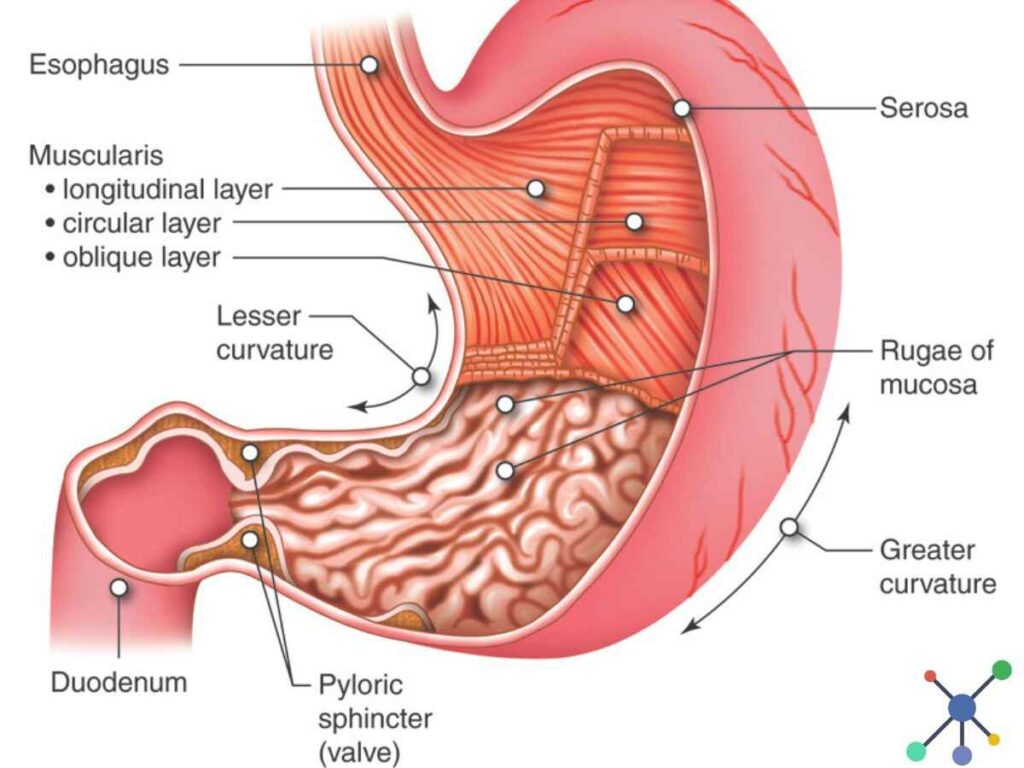
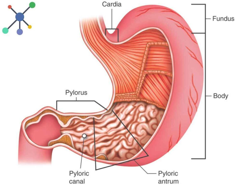
Stomach has very crucial role in the Process Of Digestion In Human Beings. Various functions of stomach are described below:
- Storage – Stomach serves as a reservoir for the food ingested. Food remains in the stomach for several hours and passes gradually into the intestine.
- Digestive – Nutritive substances undergo chemical changes by its secretory activity which provides the
enzymes; and HCI (i.e., Hydrochloric Acid) is required for the initial digestion of proteins. - HCl kills many of the ingested bacteria.
- Intrinsic factor is necessary for absorption of vitamin B12 from the small intestine. Decreased absorption of vitamin B12 produces pernicious anaemia.
- Food is released at a controlled steady rate into the duodenum to provide proper time for digestion and absorption by small intestine wall.
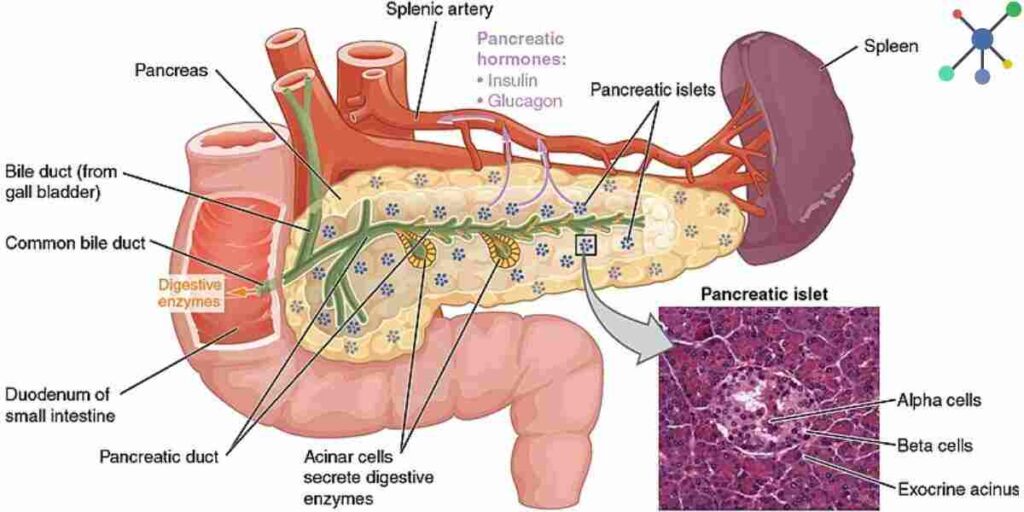
Role and Functions of Pancreas in the Process Digestion
The pancreas is a double function organ containing both ‘exocrine’ as well as ‘endocrine’ cells.
The portion of the pancreas which subserves exocrine function comprises compound alveolar tissue, that is, secretory acini and duct cells that secrete pancreatic juice.
Zymogen granules containing the digestive enzymes are concentrated at the apices of the acinar cells. The pancreatic juice is discharged from apices of the cells into the lumen of the pancreatic ducts.
The pancreatic juice passes via intercalated and excretory ducts to be collected by two ducts: duct of “Wirsung” and duct of ”Santorini”.
Pancreas secrets 1200 to 1500 ml of pancreatic juice, which is transparent, colourless fluid, isotonic with plasma.
Electrolytes present in Pancreatic Juice for Digestion for Food
Cations: K+, Na+, Ca2+, Mg2+, Zn2+
Anions: HCO3-, Cl– and traces of SO42-, HPO42-
Endocrine and Exocrine functions of Pancreas in The Process Of Digestion In Human Beings
Pancreas in human body performs both endocrine and exocrine functions. It plays a vital role in the Process Of Digestion of Food Materials In Human Beings, such as:
Exocrine Functions of Pancreas
- The release of bicarbonate: It neutralizes stomach acid by releasing bicarbonate ions into the small intestine, which in turn provides an optimal environment for enzyme function.
- Production of necessary digestive enzymes for the purpose of proper digestion: The pancreas produces the digestive enzymes lipase, which breaks down fats. It secrets another enzyme, called amylase, which breaks down carbs; and proteases, which breaks down proteins. These enzymes are secreted into the small intestine to help with the process digestion.
Endocrine Functions of Pancreas
- The pancreas secretes the hormone insulin, which aids in the uptake of glucose by cells and its subsequent storage as glycogen in the muscles and liver. This is how it help in controlling blood sugar levels.
- Production of Glucagon: In addition, it secretes glucagon, which, in the absence of insulin, stimulates the liverto release some amount of glucose into the bloodstream during periods of low blood sugar.
- Secretion of Somatostatin hormone: This hormone controls the release of insulin and glucagon, as well as blocking the production of other digestive hormones.
Functions of Liver in Human Digestive Process
1. Synthetic: Liver synthesizes
- Most of the ‘plasma proteins’ specially albumin; it does not synthesize immunoglobulins, which are synthesized in R-E system.
- Some clotting factors, e.g. fibrinogen, prothrombin and and factors of V, VII, IX and X.
- Enzymes, e.g.(i) Alkaline phosphatase (ii) SGOT (serum glutamic oxaloacetic transaminase) (iii) SGPT (serum glutamic pyruvic transaminase) (iv) SICD (sernm iso citrate dehydrogenase)
- Urea – Liver removes ammonia from the body to synthesize urea.
- Cholesterol – it is synthesized from active acetate.
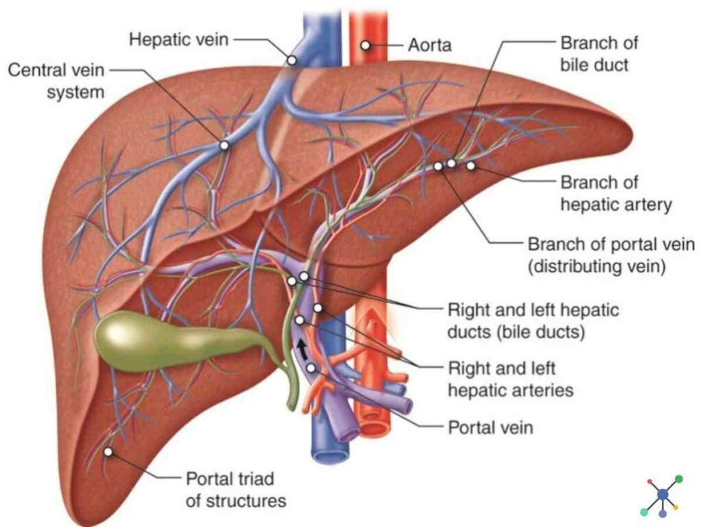
2. Metabolic
Liver is the central organ of metabolism and participates in all three major metabolisms of the body. It plays a major role in the Process Of Digestion In Human Beings.
Carbohydrate Metabolism:
Liver helps in synthesis, storage and release of glucose by the following processes:
- (i) Glycogenesis – glycogen is formed from glucose and stored in the liver.
- (ii) Glycogenolysis – liver glycogen is broken down to glucose.
- (iii) Gluconeogenesis – formation of glucose from non carbohydrate sources. (ii) and (iii) helps in regulation of blood glucose level by the liver (glucose buffer function)
Protein Metabolism:
Liver synthesizes plasma proteins, blood clotting factors, enzymes, urea and lipoprotein from ammo-acids.
Metabolism of Fats:
- (i)β-oxidation, a process which occurs within the mitochondria which oxidise the fatty acids to form active acetate i.e. aceto-acetic acid.
- (ii) Non-esterified fatty acids (NEFA) i.e. fatty acids with < 14 ‘C’ atom which reach the liver via blood are esterificd to form triglycerides in the liver.
- (iii) Synthesis of lipoproteins (e.g. HDL, LDL, VLDL, chylomicrons etc.) which are vehicles of fat.
- (iv) Synthesis of saturated fatty acids from the active acetate via Kreb’s cycle within ·the mitochondria.
- (v) Synthesis of cholesterol and phospholipids (e.g. lecithin, sphingomyelin, cephalin etc.) for cell membrane.
Process of Digestion in the Small Intestine
Carbohydrate Digestion in Small Intestine
Intestinal Juice plays a vital role in the process of digestion in the human beings. Intestinal juice contains disaccharidases i.e. enzymes for splitting disaccharides into monosaccharides, therefore:
- Sucrose, in presence of invertase (sucrase) at pH 5 to 7, gets converted into Glucose and Fructose.
- Maltose, in presence of Maltase at pH 5.8 to 6.2, gets converted into Two Molecules of Glucose.
- α-Limiting Dextrins, in presence of α-Limiting Dextrinase at pH 5.4 to 6.0, gets converted into Glucose.
Fat Digestion in Small Intestine
Intestinal lipase is particularly concerned with the ‘hydrolysis of the primary ester linkages. Triglyceride digestion begins first by removing the terminal fatty acid to produce an ‘α-β-diglyceride‘.
The other terminal fatty acid is then removed leaving a ‘β-monoglyceride‘, the main end products of fat digestion.
Protein Digestion in Small Intestine
- (i) ‘Erepsin’, a mixture of several specific enzymes, acts primarily and rapidly on peptones and polypeptides, converting them into amino-acids.
- (ii) ‘Erepsin’ can also break down ‘caseinogen’ and other proteins slowly.
- (iii) Nuclease’ and related enzymes hydrolyse the nudeic acids to liberate purine and pyrimidine bases.
Role of Large Intestine or Colon
The large intestine or colon of human digestive system extends from the cecum to the anus.
Its key tasks include completing food and water absorption, synthesising some vitamins, and forming and eliminating faeces (also known as stool).
It is about half the length of the small intestine but has a diameter of three inches, which is three times that of the small intestine.
The colon is divided into four major regions: the cecum, colon, rectum, and anal canal. The colon is further split into ascending, transverse, descending, and sigmoid colons.
Functions of Large Intestine in the process of digestion
- Further degrades food residues present inside it.
- It helps in absorption of remaining water, electrolytes, and vitamins, which were produced by enteric microorganisms present inside it.
- Propels faeces towards the rectum.
- Mucus eases the passage of excrement through the colon.
I hope the article on “Explain The Process Of Digestion In Human Beings” will help you achieve your academic goals. If you have any doubt, leave a comment below and also share with your family an friens. Thank you!
Thanks Sarbhik for such awesome content :)….. Please provide a PDF if you can…thnx again…
Thanks for your valuable comment. We are working on it. We’ll upload the PDF soon.