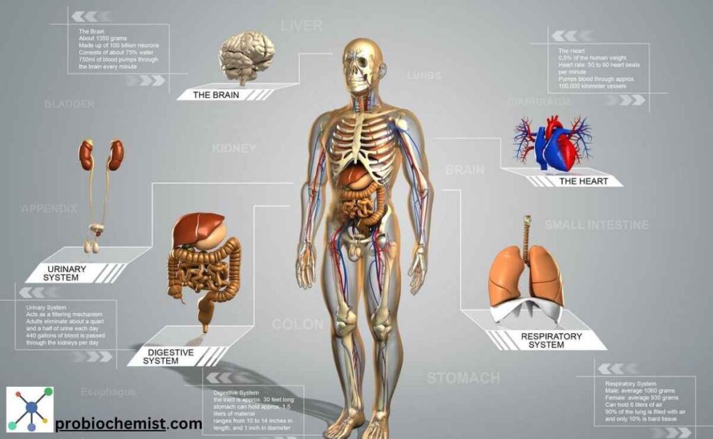
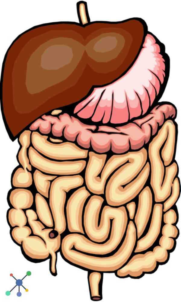
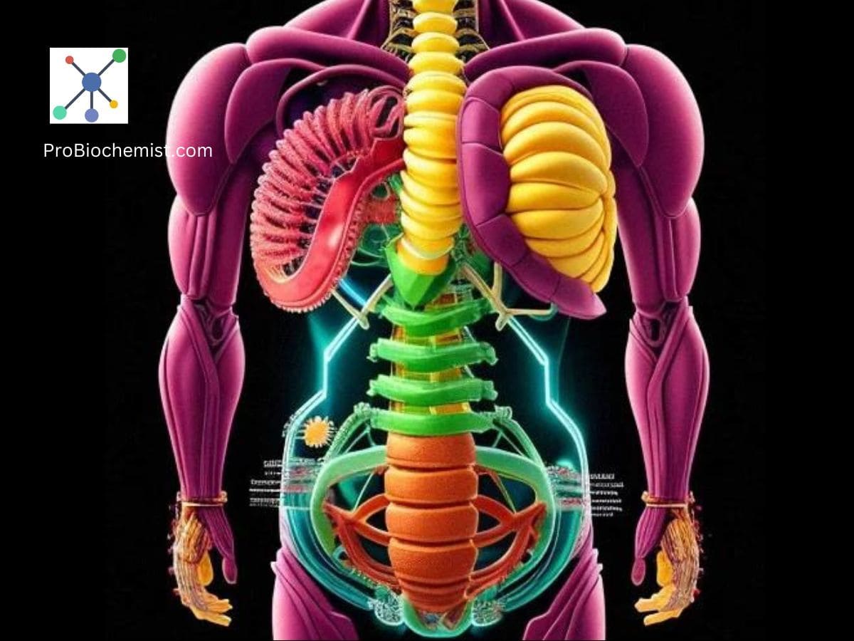
In this article, you are going to learn about how to make human digestive system model, 3D model and 3D digital model of the human digestive system in the simplest way.
Developing a handmade, realistic and precise human digestive system 3D model can be a rewarding project that provides a thorough understanding of the anatomy and operation of this complicated system.
This detailed guide will walk you through each stage of the process, from designing and acquiring materials to building and finalising your 3 dimensional model of the human digestive system.
The human digestive system is a complex network that processes food, absorbs nutrients, and eliminates waste. Building a model of this system provides a concrete picture of these processes, revealing important information about how our bodies function.

How To Make Human Digestive System 3D Model
How To Make Human Digestive System 3D Model
These following instruction will guide you how to build a 3 dimesional model that contains all of the essential components: the mouth, oesophagus, stomach, small and large intestines, and ancillary organs including the liver, pancreas, and gallbladder
- How To Make Human Digestive System 3D Model
- Building a 3D Digital Model of Human Digestive System
- The Human Digestive System Model: A Detailed Overview
Required Tools and Materials to build a 3D Digestive System Model
A. Required Materials.
1. Basic structure:
- Use foam board or cardboard as the base for your model.
- To create a 3D framework, use wire mesh or chicken wire.
- Use plastic sheets or fabric to give texture and features.
2. Digestive System Components:
- Modelling clay/playdough in different colours for organs.
- Transparent plastic tubing for the oesophagus and intestines.
- Use paper mache to create a textured surface.
- Paint: To provide realistic colours to organs.
- Use glue and tape for assembling.
3. Miscellaneous.
- Label each section of the digestive system.
- Scissors/Craft Knives: For cutting materials.
- Hot Glue Gun: Used to secure pieces.
B. Required Tools.
- Use ruler and compass for precise measurements and forms.
- Craft Knife: For precise cutting purpose.
- Use paintbrushes and markers for painting and labelling of the 3D Human Digestive System Model.

Planning the 3D Model of the Human Digestive System
A. Design Layout:
1. Determine the scale and size of your model. A 1:2 scale is a common strategy that streamlines building while keeping the human digestive model detailed enough for instructional use.
2. Organ Placement: Plan out the arrangement of the digestive organs on your basis. Place them so that it is evident how they connect and function.
B. Research:
Obtain anatomical references to assure accuracy. To comprehend each organ’s size, form, and place, consult textbooks, online resources, and anatomical charts. You might also try using digital models or apps to visualise the digestive system in 3D.
How to Build 3D model of the Human Digestive System (Using Household Items)
A. Building the Base
- To create the base, cut a wide rectangle or oval from foam board or cardboard. This will form the basis of your model.
- To support organs, design a torso-like framework with wire mesh. This is optional but useful for a three-dimensional depiction.
- Prepare the Surface: Cover the foam board with a smooth layer of paper mache or fabric, if desired. This will help the model’s texture look more realistic.
B. Making Organs
- Mouth and Esophagus: Create an open mouth with modelling clay. Form it to include teeth and a tongue. To add authenticity, paint the mouth in the right colours. Use clear plastic tubing to represent the oesophagus. Attach one end to the mouth and the other to the stomach region.
- Shape the stomach with modelling clay. It should look like a J-shaped bag. Include information such as the pyloric sphincter and stomach folds. Paint it pink and red. Position the stomach at the base, linking to the oesophagus and small intestine.
- Small Intestine: Use translucent plastic tubing to depict the small intestine. Shape it into a long, coiled structure and place it within the sculpture. [Optional: Use a thin, flexible material to simulate the villi lining the small intestine. Paint the tube to resemble the inside lining.]
- To form the large intestine, use different coloured plastic tubing or clay. It should be broader and less coiled than the small intestine. [Sections: Sculpt and place the cecum, colon (ascending, transverse, descending, sigmoid), and rectum. Connect it to your small intestine.]
- Shape the liver using clay. It should be a big, irregularly shaped organ located on the right side of the stomach. [Details: Paint the liver brownish-red and place it above the stomach. Add texture to resemble the liver’s surface.]
- Pancreas: Shape: Make a small, elongated organ from clay. [Details: Paint it pink and place it beneath the stomach, near the duodenum.]
- Gallbladder: Shape: Create a small, oval construction with clay. [Position it below the liver and paint it green to symbolise bile storage.]
- Anus: Use clay or playdough to form a little, ring-shaped structure at the end of the big intestine. [Details: Position appropriately to indicate the conclusion of the digestive tract.]
How to Assemble the 3D Model of Human Digestive System: Step by Step
1. Attach the Organs: Glue each organ to the base. Ensure that the oesophagus enters the stomach, where it links to the small intestine and ultimately the large intestine.
2. Connect Tubes: To create a more realistic representation, connect plastic tubing to show the passage from the oesophagus to the stomach and intestines.
3. Secure and Finalize: Use extra glue or tape to secure any loose parts. Ensure that all organs are properly positioned and connected.
Labelling and Detailing of the 3D Model
1. Labels: Create and attach labels to each organ. Use printed labels or stickers and place them next to the appropriate components.
2. Detailing: Enhance model realism with finishing touches like painting, shading, and texturing. You can also use arrows or other indicators to indicate the path of food through the human digestive system.
I hHope this may help you making the human digestive system model in easy way.
Additional Features Of Human Digestive System Model 3D Model
A. Functional Demonstration of the Model
1. Include interactive components, such as moveable portions or translucent sections, to demonstrate the digestive system’s structure.
2. Create educational materials, such as a handbook or digital guide, to explain the functions of each organ and digestion. Include infographics, interesting facts, and common digestive issues.
Sophisticated Features of the 3D Model
1. 3D Visualisation: To demonstrate the interior anatomy, make a 3D model with detachable pieces for a more complex project. For a more refined look, use materials like clear acrylic or plastic.
2. Digital Integration: Include QR codes or connections to online materials or films that provide a more thorough explanation of the digestive system. This can improve your model’s instructional value.
Problem-solving and upkeep
A. Typical Problems
2. Accuracy: Verify your model’s anatomical accuracy twice. Consult trustworthy sources and make any necessary modifications.
1. Stability: Verify that every component is fastened firmly. Use more glue or supports to strengthen the base and connections if the model appears unstable.
B. Upkeep
1. Handling: Take extra care when handling the model to prevent breaking any fragile pieces. To stop it from deteriorating, store it somewhere dry and cool.
2. Cleaning: Use a moist towel to gently wipe the model if it becomes dirty. Steer clear of aggressive chemicals that might harm the materials.
How to Use the 3D Model of the Human Digestive System in Education
The 3d model of the human digestive system can be used in various way, such as:
A. Use of Human Digestive System Model in the Classroom.
1. Teaching assistance: When instructing students about the digestive system, use the model as a visual assistance. It can aid pupils in comprehending the placement and purposes of each organ.
2. Interactive Lessons: Include interactive lessons in which students can follow food’s journey through the digestive system using the model and discover the functions of each organ.
B. Human Digestive System Model in Science fairs and exhibitions.
1. Presentation: – Showcase the model at scientific fairs and exhibitions. Prepare a quick presentation that describes the model’s attributes and the digestion process.
2. Engage viewers through hands-on demonstrations or quizzes about the digestive system. Provide educational resources for further study.
Building a model of the human digestive system is a fulfilling endeavor that advances knowledge of this intricate human biological system.
Through meticulous planning, building, and finishing your model, you can produce an invaluable teaching aid that illustrates the structure and operation of the digestive system.
An expertly constructed model offers a concrete depiction of how our bodies break down food and absorb nutrients, which can be used for educational purposes, scientific fairs, or just for fun. This helps people understand human physiology better.
Building a 3D Digital Model of Human Digestive System
A. Different Software Options to Make a 3D Digital Model
- Blender: sophisticated, open-source modeling software.
- Tinkercad: Easy to use and intuitive for novices.
- Fusion 360 is expert program for accurate modelling.
B. Procedure for Creating the Digital Project Model Step by Step
- Setup: Launch the 3D modeling program of your choice, then establish the project’s dimensions.
- Organ Modeling:
1. Mouth: Draw basic forms first, then add details for the tongue and teeth.
2. Model the esophagus as a tubular structure.
3. Stomach: Form an organ with a J shape and have internal folds.
4. Small Intestine: Form a structure that is coiled.
5. Large Intestine: Show a more divided, larger structure.
6. Liver: Shape this big, asymmetrical organ.
7. Pancreas: Develop a longer, smaller organ.
8. Gallbladder: Create a little oval shape model.
9. Anus: Form a structure like a ring. - Coloring and Texturing:
1. Use colors and textures to resemble real-world look.
2. Apply textures with accuracy by using UV mapping. - Assembly: Accurately place each organ.
To visualize and produce realistic visuals or animations, use rendering technologies. - Export and Share: Export in FBX, STL, or OBJ formats.
Follow these above procedure to create School project for digestive system 3d model using Household items.
The Human Digestive System Model: A Detailed Overview
The human digestive system is a fascinating and complex network that breaks down food into nutrients so the body can absorb them and be fed.
The procedure entails an intricate network of organs and processes that collaborate together to guarantee the best possible digestion and absorption.
In this article, we examine the digestive system in great detail, covering its composition, operations, and roles for each part.
1. Overview of the Digestive System
Food is broken down into smaller molecules that the body can absorb by the digestive system, which also removes waste from the body.
It starts in the mouth, travels through the stomach, small and large intestines, oesophagus, and anus before finishing at the anus. Furthermore, the liver, pancreas, and gallbladder are important auxiliary organs in the process of digestion.
A. Mouth
In the mouth, where mechanical and chemical processes take place, digestion begins. Chewing is the main mechanical process that reduces food to tiny pieces. The teeth, which have multiple shapes (incisors, canines, and molars) and are used for a variety of tasks like grinding and cutting, help with this.
Digestion depends on saliva, which is secreted by the parotid, submandibular, and sublingual salivary glands. It has the enzyme amylase, which starts the process of breaking down carbs into smaller sugars. Additionally, saliva lubricates the meal, creating a bolus that is simple to swallow.
B. Gastrointestinal Oesophagus
The oesophagus, a muscular tube that joins the throat and stomach, is where the bolus travels after leaving the mouth. The bolus is pushed downhill by peristalsis, a sequence of involuntary, wave-like muscular contractions. Mucous membranes lining the oesophagus act as lubricants and shield the lining from the corrosive effects of gastric juices.
The lower esophageal sphincter, or LES, is a valve located at the bottom of the oesophagus that keeps stomach contents from refluxing back into it.
C. Stomach
The stomach is a hollow, J-shaped organ that is in charge of additional chemical and mechanical digestion. The fundus (top portion), body (middle portion), and pylorus (bottom portion) are its three primary regions.
Gastric juices, which comprise pepsin and hydrochloric acid (HCl), are secreted by the lining of the stomach.
A pH of 1.5 to 3.5 is produced by HCl, which denatures proteins and causes pepsinogen to become pepsin, an enzyme that converts proteins into peptides. Additionally, the stomach secretes mucus to shield its lining from the acid’s corrosive effects.
Food and gastric secretions are combined in the stomach’s churning activity to create chyme, a semi-liquid material. The pyloric sphincter allows the slow release of chyme into the small intestine.
D. Small Intestine
The majority of nutritional absorption and digestion take place in the small intestine, a lengthy, coiled tube. It is split into three sections:
1. Duodenum:
The majority of chemical digestion takes place in this first part of the small intestine. It gets pancreatic juice from the pancreas and bile from the gallbladder and liver. While pancreatic enzymes (lipase, proteases, and amylase) carry out the remaining digestion of fats, proteins, and carbohydrates, bile aids in the emulsification of fats by breaking them down into smaller droplets.
2. Jejunum:
The bulk of nutrient absorption takes place in this middle portion of the small intestine. Villi and microvilli, which are microscopic projections resembling fingers that cover the jejunum’s lining, enhance the surface area available for absorption.
3. The ileum:
The last portion of the small intestine, is in charge of absorbing bile salts, vitamin B12, and any leftover nutrients. Despite having fewer villi than the jejunum, the ileum is nonetheless very effective in absorbing nutrients.
E. Pancreas
The gland that lies beyond the stomach and is responsible for both endocrine and exocrine processes is called the pancreas. The synthesis of digestive enzymes (lipase, proteases, and amylase) and bicarbonate ions is part of its exocrine action.
To help with the breakdown of proteins, lipids, and carbohydrates, the duodenum secretes these enzymes. By neutralising the stomach’s acidic chyme, bicarbonate creates the ideal pH for enzyme activity.
Though this function is distinct from digestion, the pancreas also generates the hormones glucagon and insulin, which are essential for controlling blood sugar levels.
F. Liver
The liver is a big, complex organ that is essential to digestion and metabolism. It generates bile, a chemical that facilitates fat absorption and digestion.
The gallbladder holds bile until it is needed. In addition, the liver produces the proteins required for blood coagulation, detoxifies toxic compounds, and processes nutrients taken up from the small intestine.
The liver also helps control blood sugar levels by storing excess glucose as glycogen and storing vitamins and minerals.
G. Gallbladder
Underneath the liver is a little pear-shaped organ called the gallbladder. It gathers and holds the bile that the liver produces. The gallbladder constricts and discharges bile into the duodenum through the bile ducts when fatty food enters the small intestine.
This procedure facilitates fat digestion and emulsification.
H. Large Intestine
The colon, which is part of the large intestine, is in charge of extracting water and electrolytes from the leftover indigestible food material to create solid waste (faeces). It is split up into multiple sections:
1. Cecum: The first segment of the large intestine that joins the small intestine’s ileum. It contains the appendix, a tiny, tube-shaped organ that may aid in the immune system response but has no discernible involvement in digestion.
2. Colon: The ascending, transverse, descending, and sigmoid parts make up the colon. Each area has a distinct purpose in the absorption of electrolytes, water, and certain vitamins that are produced by gut microorganisms.
3. Rectum: The last portion of the large intestine that holds on to waste materials until they are ready to be ejected is called the rectum.
I. Anus
The aperture through which excrement is released from the body is known as the anus, which is the terminal end of the digestive tract.
There are two sphincters that control it: the external anal sphincter, which is voluntary and permits conscious control over defecation, and the internal anal sphincter, which is involuntary and reacts to rectal pressure.
2. Human Digestive System Functions
The digestive system carries out a number of vital tasks:
- Ingestion: Using the mouth to take in food and liquids.
- Digestion: Using chemical and mechanical processes, food is broken down into simpler molecules throughout this process.
- Absorption: Getting nutrients into the bloodstream or lymphatic system from the digestive tract.
- Excretion: The process by which indigestible and unnecessary materials are eliminated from the body through faeces.
- Protection: Using defence systems like stomach acid and the immune system in the gut, the body guards against germs and dangerous chemicals.
3. Digestive Health and Disorders in Human Body
Maintaining gut health is critical to overall well-being. Common gastrointestinal illnesses include:
• Gastro-oesophageal reflux disease (GERD) is a disorder where stomach acid spills into the oesophagus, creating symptoms including heartburn.
• Peptic ulcers are sores on the stomach, small intestine, or oesophagus that can be caused by H. pylori infection or NSAID use.
• IBS is a functional gastrointestinal illness characterised by abdominal pain, bloating, and changes in bowel patterns.
• IBD, which includes Crohn’s disease and ulcerative colitis, is characterised by persistent inflammation of the digestive tract.
• Coeliac disease is an autoimmune illness where gluten causes damage to the small intestine.
The human digestive system is a complex and efficient system that processes food, absorbs nutrients, and eliminates waste. It entails a number of organs and systems that work together to ensure the body receives the nutrition it requires while remaining healthy.
Understanding the digestive system’s anatomy and function can help you recognise and treat digestive diseases, as well as promote overall digestive health.
This post will help you to create a perfect handmade digestive system 3d model project for your school, college or university class. If you find this post on ‘How To Make Human Digestive System Model‘ helpful, please share this article and also leave a comment. Thank you!

Thank you admin for such helpful content… I just made a 3d model of human digestive system and submitted to the school and got excellent marks…. Thank you again!
You are welcome… I appreciate you taking the time to leave a comment…